
Clinical Leaders & Researchers
Associated Projects: Advanced imaging biomarkers
Dr. Jay Detsky is a radiation oncologist at the Odette Cancer Centre at Sunnybrook Health Sciences Centre in Toronto. He specializes in treating CNS malignancies and prostate cancer. He also has a PhD in Medical Biophysics with an expertise in magnetic resonance imaging.
Dr. Detsky’s research interests include advanced imaging biomarkers to help not only diagnose cancers, but to help with radiation treatment planning, and to predict response to therapy. His contribution to the IGAR project is to aid with the clinical pathway for men from the diagnosis through treatment of prostate cancer to help identify the best use of an autonomous medical robot to facilitate prostate cancer therapy.
Associated Projects: AI in Breast Imaging
Dr. Curpen has been practicing as a radiologist specializing in Breast Imaging for the past 26 years in an academic center. Her current research program is focused on improving the diagnostic accuracy of ultrasound and magnetic resonance imaging using quantitative imaging analyses as well as investigating AI in Breast Imaging. The primary endpoint is to provide better imaging tools to radiologists and other clinicians. The work on quantitative ultrasound for treatment response monitoring has already led to several publications (Sannachi et al. 2018; Sadeghi-Naini et al. 2017; Tran et al. 2017) and currently co-leading research on the use of quantitative magnetic resonance imaging to detect biological changes in breast tumors treated with chemotherapy.
Dr. Curpen was also involved with a study to determine whether women with dense breasts and DCIS have a higher risk of recurrence. The work will ultimately lead to improved outcomes for patients by tailoring treatments according to imaging signatures obtained from quantitative analyses. Sunnybrook was a pioneer in MRI screening for women at high risk of developing breast cancer and our screening program predates the Ontario-wide high-risk breast screening program which was introduced in 2011. As well as contributing to studies on survival of MR screened patients (Passaperuma et al. 2012), she obtained funding to carried out a study to determine whether MR screening was beneficial in women with dense breasts and a previous breast cancer (Nadler et al. 2017). This group is not currently covered by the OBSP high risk program but results suggest that the addition of MRI screening should be considered for surveillance of women with a combination of these risk factors, particularly if they have a family history of breast cancer and are not on anti-estrogen therapy. If this finding is reproduced in a larger scale clinical trial it could have a major impact on breast screening protocols.
She has worked to improve breast screening using mammography and am involved with TMIST, which is a large 3-year study multi-institutional study to evaluate tomosynthesis for screening. More than 8000 women will take part in 3 sites for up to 3 years to evaluate. At Sunnybrook Health Sciences Centre present, 2000 have been recruited.
Another big part of our practice is to do preoperative localization of non-palpable breast cancers. A novel way of localization is to use radioactive seeds preoperatively. I recently published a paper on the cost of preoperative localization by radioactive seeds versus wire localization.
Associated ProjectS: 3D ultrasound in image-guided interventions
Dr. Aaron Fenster’s area of research is focused on the use of 3D ultrasound in image-guided interventions. He is a Scientist at the Robarts Research Institute, founder and past Director of the Imaging Research Laboratories, Chair of the Division of Imaging Sciences of the Department of Medical Imaging at The Western University, founder and past Director of the graduate Program in Biomedical Engineering, Director Imaging Program, of the Ontario Institute for Cancer Research, Founder, and Centre Director of Centre for Imaging Technology Commercialization. He has published 350 peer-reviewed papers and has 52 patents filed or issued. He is a Fellow of CAHS, IEEE, SPIE, COMP, IOMP, AAPM and CCPM. He held a Canada Research Chair-Tier 1 in Biomedical Engineering from 2000 to 2014. He is the first recipient of the Premier’s (Ontario) Discovery Award for Innovation and Leadership (2007), the Hellmuth Prize for Achievement in Research at the UWO (2008), and the Canadian Organization of Medical Physicists (COMP) Gold Medal Award (2010). In 2013, the International Organization for Medical Physics (IOMP) at its annual meeting named Fenster as one of the 50 international Medical Physicists who have made an outstanding contribution to the advancement of Medical Physics over the past 50 years. In 2020, he received the Order of Ontario.
Associated Projects: Breast Pathology
Fang-I Lu completed undergraduate medical education at McGill University Faculty of Medicine, Montreal, Canada from 2002-2006, followed by Anatomical Pathology Residency Program at University of British Columbia, Vancouver Canada from 2006-2011 and a Breast Pathology Fellowship at Memorial Sloan-Kettering Cancer Centre, New York, USA from 2011-2012.
She is currently a staff pathologist at Sunnybrook Health Sciences Centre, Toronto, Canada, and an assistant professor at University of Toronto. Her area of clinical expertise is in breast pathology and she also actively participates in postgraduate and continuing medical education at University of Toronto.
Fang-I Lu is the pathology lead in CSii and her roles include 1) reviewing pathology aspect of grant applications, 2) investigating laboratory methods to allow for rapid tissue processing and 3) providing breast pathology expertise for the development of artificial intelligence and machine learning in breast pathology.
Dr. Susan Reid is Professor and Chair of the Academic Department of Surgery at McMaster University, where she also completed her own medical training. The first female appointed Chair of a surgical department in a Canadian medical school, she heads a group of over 300 surgeons and 180 residents/fellows.
Dr. Reid has worked as a general surgeon, trauma surgeon, and intensivist during her more than 26 years with Hamilton Health Sciences, and co-founded the Greater Hamilton Area (GHA) Surgical Centre. With a special interest in diseases of the breast, she continues to champion and participate in research designed to improve patient care, outcomes, and quality of life for those with breast cancer.
She has served as president of the Canadian Association of General Surgeons, and is a dedicated teacher, mentor, and researcher in surgical education.
Dr. Michael D. Noseworthy, PhD, PEng (LEL). Received a M.Sc. from the University of Guelph for work in the evaluation of anaesthetic hepatotoxicity using MRI, transmission electron microscopy (TEM) and in vivo 31P-NMR. Obtained a PhD from University of Guelph (1997) specializing in applications of MRI/MRS and electron paramagnetic resonance (EPR) methods to assess free radical induced brain damage. From 1997-1999 was a postdoctoral fellow in Imaging Physics, Sunnybrook Health Sciences Centre (Toronto) working on the evaluation of tissue microvasculature through development of correlative MRI and energy dispersive X-ray microanalysis (EDXS). In January 2000 to August 2003 worked as an MRI physicist at The Hospital for Sick Children and University Health Network (UHN), Toronto, and was Assistant Professor in Medical Biophysics and Medical Imaging, University of Toronto. He was recruited to St. Joseph’s Healthcare and Brain-Body Institute, McMaster University in August 2003. Following 3 years as an Assistant Professor in Radiology and Medical Physics at McMaster, Dr. Noseworthy obtained a tenure-track assistant professor position in Electrical & Computer Engineering at McMaster University, where he currently resides as a full professor. He was the Co-Director of the School of Biomedical Engineering (from 2010 to 2020) and has been Director of Imaging Physics and Engineering at the Imaging Research Centre, St. Joseph’s Healthcare, Hamilton since 2003. Dr. Noseworthy also has Special Professional Staff status at St. Joseph’s Healthcare in both Radiology and Nuclear Medicine. He has received funding from numerous sources including NSERC, CIHR, CFI and ORF, has published almost 150 peer reviewed papers, 260 conference papers and had supervised almost 70 graduate students and postdocs. Dr. Noseworthy is Co-Founder and Director of TBIfinder, a data analytics company specializing in brain injuries. Dr. Noseworthy is a member of the Professional Engineers of Ontario (PEO), Institute of Electrical and Electronics Engineers (IEEE), International Society for Magnetic Resonance in Medicine (ISMRM) and European Society for Magnetic Resonance in Medicine and Biology (ESMRMB).
Dr Andrew Loblaw is a Radiation Oncologist, Clinician Scientist, and dual Professor in the Department of Radiation Oncology and the Institute of Health Policy Management & Evaluation at the University of Toronto.
He received a Bachelor of Science in Physics from the University of British Columbia and his Doctor of Medicine from Queen’s University. He completed his specialty training in Radiation Oncology concurrent with a Masters degree in Clinical Epidemiology to graduate from Royal College’s Clinician Investigator Program all at the University of Toronto.
Dr Loblaw’s clinical practice and research interest focus on improving outcomes for men with prostate cancer and the healthcare system. He has a particularly interest in the design and conduct of clinical trials, the generation and dissemination of evidence-based guidelines and in image-guided radiotherapy.
Dr Loblaw is an Ontario Association of Radiation Oncology Clinician Scientist and a Senior Scientist at the Sunnybrook Research Institute. A Fellow of the American Society of Clinical Oncology (FASCO), he was previous Co-Chair of the ASCO’s Genitourinary Advisory Group and remains Co-Chair of the GU group for Cancer Care Ontario’s Program in Evidence-Based Care. He has authored over 225 peer-reviewed papers and has been awarded grant funding of over $37M.
Kathy Schilling, MD is a board-certified diagnostic radiologist specializing in breast imaging who has spent the last 30 years of her career at Boca Raton Regional Hospital in Boca Raton, Florida. She is Medical Director of the Christine Lynn Women’s Health and Wellness Institute. The Institute is nationally and internationally renowned to provide the most comprehensive breast imaging and interventional services for the women in South Florida. Dr. Schilling has grown the program to employ 8 dedicated breast imagers performing >120,000 procedures per year and identifying >650 new breast cancers on an annual basis. Her area of greatest interest over the years is identifying and validating new imaging and interventional techniques. In the early 1990’s she was an early adopter of percutaneous breast biopsy per-formed with stereotaxis and ultrasound. Over the years she has taught many radiologists and surgeons these techniques which expanded in the more recent years to include MRI and PEM guided biopsy procedures. Dr. Schilling was involved in the validation process of the ICAD computer aided detection device in 2000, Positron Emission Mammography in 2006, Automated 3D Breast Ultrasound, Hologic Digital Breast Tomosynthesis, Shear Wave Elastography and Optoacoustic Imaging of the breast. She authored and co-authored many peer review articles and has spoken widely on her experience nationally and internationally. She has developed several in-house fellowship programs where physicians spend time at the Center for Breast Care learning these sophisticated technologies and the successful efficient functioning of a comprehensive breast center.
Dr Schilling attended medical school at the University of Miami and did an internship in Internal Medicine at Jackson Memorial Hospital in Miami. This was followed by a four-year residency in Diagnostic Radiology at Mt. Sinai Medical Center in Miami Beach and an additional fellowship year in Diagnostic Imaging. Dr. Schilling is a partner in Boca Radiology Group, PA., a 36-person radiology group in Boca Raton, Florida.
Dr. Tran is an RT clinician scientist in the Department of Radiation Oncology at the Odette Cancer Centre, and Assistant Professor of Radiation Oncology at the University of Toronto. He is also appointed as scientist at Sunnybrook Research Institute. Dr. Tran’s laboratory is developing a computational oncology imaging program with a focus on predictive and prognostic modelling in high-risk breast cancer using digital pathology imaging biomarkers.
Dr. Tran is also a senior research fellow at Sheffield Hallam University (Sheffield, United Kingdom), and a member of the University of Toronto Centre for AI Research and Education in Medicine.
Dr. Nathalie Duchesne has been working in breast imaging and intervention since 1996, is now Breast radiologist at Hopital du Saint-Sacrement in Quebec City. She is Academic Clinical Associate Professor at Universite Laval in Quebec City.
Nathalie received her Medical Doctorate in 1990 and her Diagnostic Radiology postgraduate degree in 1995, both from University Laval, Quebec City. She has performed rotations in university hospitals both in Australia and The Netherlands, and worked on a fellowship program in interventional MRI and bone tumors at Harvard University. The latter was completed in breast imaging at the Universite de Montreal. She also holds a B.Sc. degree in Biology.
Dr. Duchesne's main clinical and research interests include breast biopsy tool development, minimally-invasive therapy, as well as new types of breast imaging and cancer detection. She is a pioneer in vacuum-assisted breast biopsy, having done many world and Canadian premieres for various devices. She is an internationally known speaker having given numerous national and international conferences, with a track record of publications in the areas of breast imaging and intervention. She has lectured in the Breast Imaging and Intervention Series (2001-2003). She is a member of various international scientific societies, and has received many awards from her peers, such as the Young Radiologist Investigator Award of the Year for 2005 by the Canadian Association of Radiologists, and the 2008 Personality of the Year in Radiology from the Société Canadienne-Française de Radiologie / Association des Radiologistes du Québec for her personality, scientific contribution, and humanitarian work.
Dr. Nathalie Duchesne is the founder and Director of The Breast Practices, organizing the now world famous interdisciplinary The Breast Course and The Breast Days. Through these courses, more than 2,000 physicians from 61 countries have received teaching, contributing to the improvement of breast and women's health worldwide.
Associated Projects: Seed Localization Project
Although minimally invasive techniques are the preferred route for diagnosis, there are some cases where minimally invasive techniques cannot be used, and surgical removal is the only way to proceed. In cases where a lesion cannot be located by palpation (feeling of a physical "hard" lump) then the surgeon has to be guided to the correct location. This is typically done using very fine "hook wires" that are placed using x-ray or ultrasound directly into the breast where the anomaly is prior to surgery.
Dr Peter Lovrics (Head of General Surgery) has performed a clinical trial of a new technique called seed localization where a very small radioactive "seed" (smaller than a grain of rice) is placed near the area of interest prior to surgery (see Figure 1). In the operating theatre Dr Lovrics can locate the seed using a radioactive detector, and hence identify the lesion, which can be removed with much greater accuracy than the conventional hook-wire procedure. This has been proven to greatly reduce the possibility of follow up surgery. It also allows the surgeon greater flexibility in their angle of approach to access the tumor (allowing better cosmesis) and can result in a smaller sample needing to be taken (greater accuracy).
With IGAR we have the ability to make the next step and use MRI guidance to place the seed in certain types of cancer that are invisible to ultrasound and x-ray
mammography. We believe this will play a vital role for surgical guidance in excisional biopsy and breast conserving lumpectomy procedures.
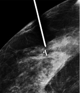
figure 1. A radioactive seed being placed under x-ray guidance
Kathy Schilling, MD is a board-certified diagnostic radiologist specializing in breast imaging who has spent the last 30 years of her career at Boca Raton Regional Hospital in Boca Raton, Florida. She is Medical Director of the Christine Lynn Women’s Health and Wellness Institute. The Institute is nationally and internationally renowned to provide the most comprehensive breast imaging and interventional services for the women in South Florida. Dr. Schilling has grown the program to employ 8 dedicated breast imagers performing >120,000 procedures per year and identifying >650 new breast cancers on an annual basis. Her area of greatest interest over the years is identifying and validating new imaging and interventional techniques. In the early 1990’s she was an early adopter of percutaneous breast biopsy per-formed with stereotaxis and ultrasound. Over the years she has taught many radiologists and surgeons these techniques which expanded in the more recent years to include MRI and PEM guided biopsy procedures. Dr. Schilling was involved in the validation process of the ICAD computer aided detection device in 2000, Positron Emission Mammography in 2006, Automated 3D Breast Ultrasound, Hologic Digital Breast Tomosynthesis, Shear Wave Elastography and Optoacoustic Imaging of the breast. She authored and co-authored many peer review articles and has spoken widely on her experience nationally and internationally. She has developed several in-house fellowship programs where physicians spend time at the Center for Breast Care learning these sophisticated technologies and the successful efficient functioning of a comprehensive breast center.
Dr Schilling attended medical school at the University of Miami and did an internship in Internal Medicine at Jackson Memorial Hospital in Miami. This was followed by a four-year residency in Diagnostic Radiology at Mt. Sinai Medical Center in Miami Beach and an additional fellowship year in Diagnostic Imaging. Dr. Schilling is a partner in Boca Radiology Group, PA., a 36-person radiology group in Boca Raton, Florida.
Dr. Tran is an RT clinician scientist in the Department of Radiation Oncology at the Odette Cancer Centre, and Assistant Professor of Radiation Oncology at the University of Toronto. He is also appointed as scientist at Sunnybrook Research Institute. Dr. Tran’s laboratory is developing a computational oncology imaging program with a focus on predictive and prognostic modelling in high-risk breast cancer using digital pathology imaging biomarkers.
Dr. Tran is also a senior research fellow at Sheffield Hallam University (Sheffield, United Kingdom), and a member of the University of Toronto Centre for AI Research and Education in Medicine.
Dr. Nathalie Duchesne has been working in breast imaging and intervention since 1996, is now Breast radiologist at Hopital du Saint-Sacrement in Quebec City. She is Academic Clinical Associate Professor at Universite Laval in Quebec City.
Nathalie received her Medical Doctorate in 1990 and her Diagnostic Radiology postgraduate degree in 1995, both from University Laval, Quebec City. She has performed rotations in university hospitals both in Australia and The Netherlands, and worked on a fellowship program in interventional MRI and bone tumors at Harvard University. The latter was completed in breast imaging at the Universite de Montreal. She also holds a B.Sc. degree in Biology.
Dr. Duchesne's main clinical and research interests include breast biopsy tool development, minimally-invasive therapy, as well as new types of breast imaging and cancer detection. She is a pioneer in vacuum-assisted breast biopsy, having done many world and Canadian premieres for various devices. She is an internationally known speaker having given numerous national and international conferences, with a track record of publications in the areas of breast imaging and intervention. She has lectured in the Breast Imaging and Intervention Series (2001-2003). She is a member of various international scientific societies, and has received many awards from her peers, such as the Young Radiologist Investigator Award of the Year for 2005 by the Canadian Association of Radiologists, and the 2008 Personality of the Year in Radiology from the Société Canadienne-Française de Radiologie / Association des Radiologistes du Québec for her personality, scientific contribution, and humanitarian work.
Dr. Nathalie Duchesne is the founder and Director of The Breast Practices, organizing the now world famous interdisciplinary The Breast Course and The Breast Days. Through these courses, more than 2,000 physicians from 61 countries have received teaching, contributing to the improvement of breast and women's health worldwide.
Associated Projects: Seed Localization Project
Although minimally invasive techniques are the preferred route for diagnosis, there are some cases where minimally invasive techniques cannot be used, and surgical removal is the only way to proceed. In cases where a lesion cannot be located by palpation (feeling of a physical "hard" lump) then the surgeon has to be guided to the correct location. This is typically done using very fine "hook wires" that are placed using x-ray or ultrasound directly into the breast where the anomaly is prior to surgery.
Dr Peter Lovrics (Head of General Surgery) has performed a clinical trial of a new technique called seed localization where a very small radioactive "seed" (smaller than a grain of rice) is placed near the area of interest prior to surgery (see Figure 1). In the operating theatre Dr Lovrics can locate the seed using a radioactive detector, and hence identify the lesion, which can be removed with much greater accuracy than the conventional hook-wire procedure. This has been proven to greatly reduce the possibility of follow up surgery. It also allows the surgeon greater flexibility in their angle of approach to access the tumor (allowing better cosmesis) and can result in a smaller sample needing to be taken (greater accuracy).
With IGAR we have the ability to make the next step and use MRI guidance to place the seed in certain types of cancer that are invisible to ultrasound and x-ray
mammography. We believe this will play a vital role for surgical guidance in excisional biopsy and breast conserving lumpectomy procedures.
figure 1. A radioactive seed being placed under x-ray guidance
Julian Dobranowski continues to serve as the Clinical Lead for the IGAR Breast Investigation Team. Dr. Dobranowski's extensive experience in diagnostic imaging has proved critical in the correct definition of patient care pathways and clinical requirements for the breadboard prototype. Under his expert direction, CSii engineering efforts have enjoyed greater access to clinical expertise and quick clinical feedback, which have been paramount to the ongoing success of the program. His involvement has also allowed CSii to quickly access top diagnostic imaging experts throughout the province who have helped to shape the direction of the program with a broader view of clinician uses and requirements for the IGAR breast system.
Dr. Boylan is Chief of Dept Diagnostic Imaging at St. Joseph's Healthcare Hamilton. He graduated from Medicine at University College Dublin and underwent his postgraduate training in Diagnostic Imaging at the University of Manchester in the United Kingdom. Dr. Boylan underwent further fellowship training in Breast Imaging, Chest Imaging, and Abdominal Imaging at Sunnybrook and UHN. His research interests include quantitative CT, new MRI techniques and image-guided procedures.
Dr. Ananth Ravi is also a board-certified medical physicist as well as an associate professor in the department of radiation oncology at the University of Toronto. Dr. Ravi has considerable clinical and research experience in both image-guided brachytherapy as well as improving guidance for surgical oncology. Dr. Ravi is one of the founders and is the Chief Scientific Officer for MOLLI Surgical, a company focused on making precision surgery simpler through the incorporation of innovative and robust tracking technologies.
Dr. Calvin Law, MD, MPH, FRCSC, is also a cancer surgeon specializing in hepatobiliary, pancreatic and gastrointestinal surgical oncology.
As a leader, Dr. Law has held several leadership positions prior to his current role including being the Cancer Care Ontario Head and Regional Lead of Surgical Oncology for Toronto Central North and the Chair for Gastrointestinal Oncology Site Group at the Edmond Odette Cancer Centre. He was also the co-founder of the Susan Leslie Clinic for Neuroendocrine Tumours.
From an academic point of view, Dr. Law holds the rank of Full Professor at the University of Toronto in the Department Surgery and the Department of Health Policy, Management and Evaluation. He also serves as an Affiliate Scientist at Sunnybrook Research Institute and has served as an Adjunct Scientist for the Institute of Clinical Evaluative Sciences. Dr. Law was the inaugural holder of the Sherif and Mary-Lou Hanna Research Chair in Surgical Oncology at the University of Toronto. He has also completed a term as a Career Scientist for the Ministry of Health and Long-Term Care of Ontario. His research focuses on health services research and population outcomes, where he has published over 160 peer reviewed publications.
As an educator, Dr. Law has been recognized with teaching awards at the undergraduate, graduate and post-graduate levels, including the Canadian Association of General Surgery Resident Teaching Award, the Bruce Tovee Surgical Teaching Award and the Robert Mustard Mentorship Award. He has also served a term as an Examiner for the Royal College of Physicians and Surgeons of Canada.
Dr. Law completed his medical school training at the University of Toronto, after which time he travelled to McMaster University to complete his General Surgery training. He returned to the University of Toronto to complete his Surgical Oncology Fellowship training and went on to Cambridge, Massachusetts to complete his Masters of Public Health at Harvard University. He then returned to Canada, and settled at the University of Toronto, and Sunnybrook Health Sciences Centre.
Qiyin Fang is a professor of Engineering Physics at McMaster University and held the Canada Research Chair in Biophotonics from 2005-2016. Dr. Fang is an elected fellow of the International Society for Optical Engineering (SPIE).
Dr. Fang's current research interests include multimodality sensing and imaging technologies for biomedical applications, e.g. optical biopsy, wide-field imaging, endoscopy, microscopy, point-of-care/in-home/wearable sensing systems, and distributed environmental sensing. Prior to his current position at McMaster, Dr. Fang was a research scientist in the Minimally Invasive Surgical Technology Institute at the Cedars-Sinai Medical Center in Los Angeles.